
Pars intermedia
| Pars intermedia | |
|---|---|
 Median sagittal through the hypophysis of an adult monkey. (Pars intermedia labeled at bottom center.) | |
| Details | |
| Identifiers | |
| Latin | pars intermedia adenohypophyseos |
| TA98 | A11.1.00.004 A09.4.02.017 |
| TA2 | 3858 |
| TH | H3.08.02.2.00007 |
| FMA | 74632 |
| Anatomical terminology | |

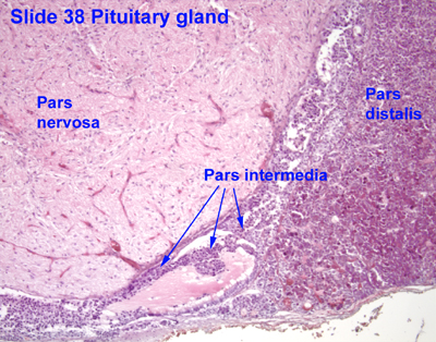
Pars intermedia is the portion of the pituitary forming the boundary between its anterior lobe and its posterior lobe.[1] The pars intermedia secretes α-melanocyte-stimulating hormone (α-MSH), and corticotropin-like intermediate peptide. In humans, it does not appear to be functionally significant and is normally small, except during foetal life and pregnant women (in whom it may be responsible for increased skin pigmentation). It appears to be tonically inhibited by the hypothalamus.
It is well developed in some animals like amphibians[2] in whom it mediates active camouflage, causing darkening of the skin when placed against a darker background.
Anatomy
[edit]Microanatomy
[edit]It contains colloid-filled cysts and two types of cells - basophils and chromophobes. The cysts are the remainder of Rathke's pouch. As technically part of the anterior pituitary, it separates the posterior pituitary and pars distalis. It is composed of large, pale cells that encompass the aforementioned colloid-filled follicles.[1]
Physiology
[edit]The pars intermedia appears to be tonically inhibited by stimuli from the hypothalamus (either by dopaminergic innervation or by vascular mechanism) as experimental sectioning of the pars intermedia from the hypothalamus has been noted to result in hypertrophy of the pars intermedia in various animals.[2]
Function
[edit]The pars intermedia is responsible for secreting α-melanocyte-stimulating hormone, and corticotropin-like intermediate peptide.[2]
It is prominent only during foetal life and in pregnant women, but is otherwise negligible. The characteristic pattern of skin hyperpigmentation seen during pregnancy may be a result of increased circulating maternal a-MSH (which may have originated from either the maternal or foetal pars intermedia), but a-MSH secretion does not seem to be involved in skin tanning in response to light exposure.[2]
Other animals
[edit]In lower vertebrates (fish, amphibians), MSH from the pars intermedia is responsible for darkening of the skin, often in response to changes in background color.[citation needed] This color change is due to MSH stimulating the dispersion of melanin pigment in the animal's skin melanocyte chromatophores. Some animals will thus increase a-MSH secretion when placed against a dark background[2] as a means of active camouflage.
See also
[edit]References
[edit]- ^ a b Ilahi, Sadia; Ilahi, Tahir B. (3 October 2022). "Anatomy, Adenohypophysis (Pars Anterior, Anterior Pituitary)". Anatomy, Adenohypophysis (Pars Anterior, Anterior Pituitary). StatPearls Publishing. Retrieved 9 February 2023.
- ^ a b c d e Davies, Andrew; Blakeley, Asa G. H.; Kidd, Cecil, eds. (2001). Human Physiology. pp. 397–398. ISBN 978-0-443-04559-2.
External links
[edit]- Histology image: 14001loa – Histology Learning System at Boston University
- Histology image: 14101loa – Histology Learning System at Boston University
- Histology image: 38_11 at the University of Oklahoma Health Sciences Center
- UIUC Histology Subject 991